Lyophilised Peptides, Reconstitution & −20 °C Storage: A Practical Guide to the Corex Vitality™ Learning Hub
Lyophilised Peptides, Reconstitution & −20 °C Storage: A Practical Guide to the Corex Vitality™ Learning Hub
For in-vitro laboratory research use only. Not for human or veterinary use.
Quick summary
Research peptides are supplied freeze-dried (lyophilised) because they stay stable far longer as a dry solid than in liquid.
A small white cake or loose powder at the bottom of the vial is normal, and so is a vial that looks almost empty.
Choose the solvent from the peptide’s sequence: count its basic and acidic groups, test a small portion first, and use oxygen-free solvent for sequences containing Cys, Met or Trp.
Concentration is simple maths: vial mass (mg) ÷ solvent volume (mL). Our free calculator on the Learning Hub does it for you.
Store dry vials at −20 °C or colder for longer storage (2–8 °C is fine short-term; room-temperature transit is normal). Split reconstituted stock into single-use aliquots, freeze them and thaw each only once.
Reconstituting and storing a lyophilised peptide correctly is one of the simplest ways to protect the quality of your in-vitro work. This guide explains what happens at each stage in plain English, and shows how to use the new interactive tools in the Corex Vitality™ Learning Hub to check your numbers before you pick up a pipette.
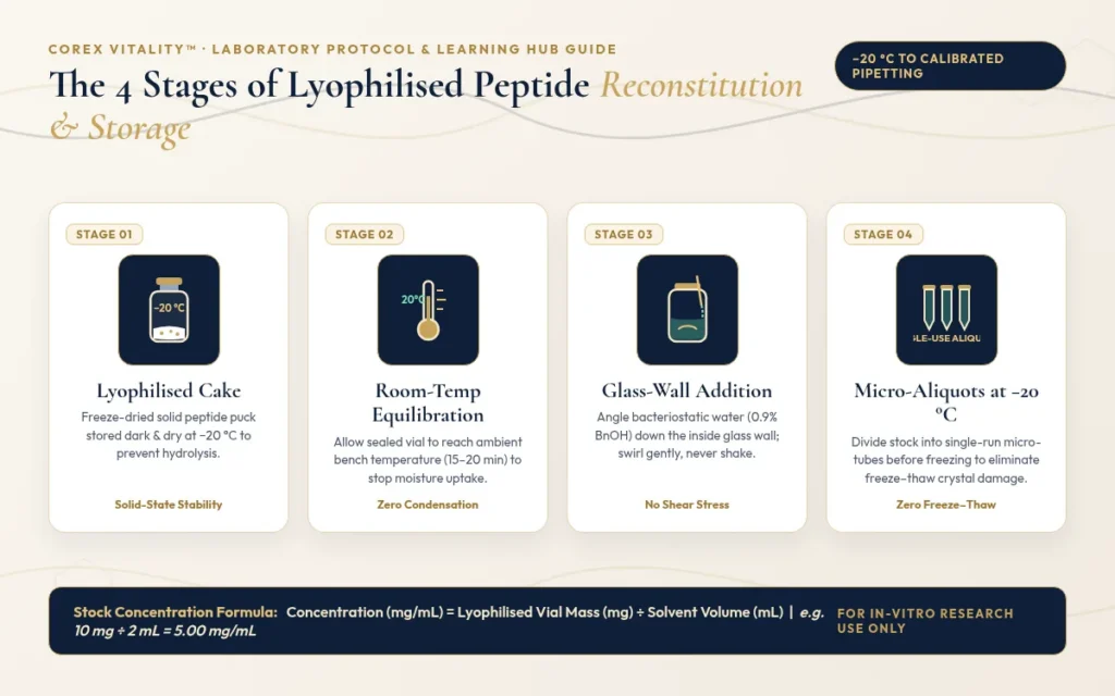
Simplified overview of the four stages that protect a research peptide from vial to aliquot.
1. How to use the Corex Vitality™ Interactive Learning Hub
Our Learning Hub now opens with three interactive tools, all in one panel near the top of the page.
Interactive Knowledge & Compound Explorer. Type a peptide name, CAS number or topic into the search bar, or tap a pathway pill such as “Storage & Reconstitution”. Compound cards show the CAS number, molecular weight, sequence or formula and storage temperature, with one-tap links to the product page and the certificate of analysis. New guides, including this one, appear automatically.
In-Vitro Laboratory Reconstitution & Dilution Calculator. Pick the vial mass, diluent volume and target aliquot, or type your own values. The calculator shows the stock concentration in mg/mL (and µg/mL), the micropipette volume per aliquot in mL and µL, and how many aliquots one vial gives. A vial diagram fills as you change the volume.
4-Step Lab Protocol & COA Walkthrough. Four clickable steps cover receiving and storage, reconstitution, reading an HPLC and mass spectrometry certificate, and aliquoting. Each step ends with a short self-check question and a link to verify your batch.
2. Why research peptides arrive as a freeze-dried “puck”
Lyophilisation, or freeze-drying, removes water from a frozen peptide solution by sublimation: under a deep vacuum at low temperature, ice turns straight into vapour without melting. What is left is a dry, porous solid.
This matters because water drives most of the ways a peptide breaks down. In solution, peptide bonds can be slowly cut by hydrolysis, and residues such as methionine and cysteine can oxidise. At room temperature that can become measurable within days or weeks. As a dry solid, the same molecule is far more stable.
The result is usually a small white cake, sometimes called a puck, or a loose powder at the bottom of the vial. A 2 mg or 5 mg vial can look almost empty, and the cake may crack or shrink in transit. None of this affects the contents: the quantity in the vial is measured on the certificate of analysis.
3. Choosing the right laboratory solvent
There is no single solvent that suits every peptide. The best starting point depends on the sequence, so work it out before you open the vial and always test a small portion first, keeping the rest dry.
Step 1: estimate the overall charge
Count the basic groups (lysine K, arginine R, histidine H and a free N-terminus) and the acidic groups (aspartic acid D, glutamic acid E and a free C-terminus). An acetylated N-terminus or an amidated C-terminus does not count. This is only an approximate guide, and lipidated peptides often behave differently.
More basic groups (basic peptide): try water first. If it is slow, add a small amount of dilute acetic acid, then dilute to volume.
More acidic groups (acidic peptide): try water or a neutral buffer first. If it is slow, add a small amount of dilute (about 0.1%) aqueous ammonia, then dilute to volume.
Around neutral pH: phosphate-buffered saline (PBS) at pH 7.0–7.4 often works for concentrations up to about 1 mg/mL.
Very hydrophobic or neutral sequences: dissolve in a small volume of an organic solvent such as DMSO, DMF or acetonitrile, then add the aqueous buffer slowly while mixing. Check that the organic solvent is compatible with your assay.
Step 2: check for sensitive residues
Sequences containing cysteine, methionine or tryptophan oxidise easily, so use freshly degassed, oxygen-free solvent and keep air exposure to a minimum. Do not add reducing agents such as DTT to peptides that rely on a disulfide bridge. Asparagine and glutamine shorten shelf life in solution, so prepare these fresh and aliquot promptly. Each Corex Vitality™ product page has a Storage & Handling box with a compound-specific note.
Step 3: water, sterile water or bacteriostatic water
Bacteriostatic water is sterile water containing 0.9% benzyl alcohol. The benzyl alcohol slows bacterial growth but does not sterilise, it is not suitable for every peptide, and it can interfere with cell-based assays. Our BAC Water for laboratory reconstitution is one option. Sterile water or a sterile buffer without preservative is the better choice for a single session or for assays where benzyl alcohol could interfere.
Swirl gently, and if needed use brief sonication in a water bath. Do not heat the solution. The solubility approach above is paraphrased in our own words from Bachem’s peptide solubility guidelines.
4. Step-by-step reconstitution and concentration maths
Let the vial reach room temperature. Opening a cold vial draws moisture from the air onto the dry powder as condensation. Leave it sealed, ideally in a desiccator, until it reaches room temperature. Then weigh out or reconstitute quickly and reseal tightly. Peptide powders absorb moisture, and absorbed water lowers the real peptide content of what you weigh.
Add solvent down the glass wall. Aim the stream at the inside wall of the vial, never directly onto the cake, and add it slowly.
Swirl gently. Roll or swirl the vial until the solution is clear. Do not shake or vortex: vigorous mixing creates air–liquid surfaces where peptides can unfold and clump together.
Label it with the compound, concentration, date and batch number.
Worked example: a 10 mg vial with 2 mL of solvent gives 10 ÷ 2 = 5.00 mg/mL, which is 5,000 µg/mL. A 500 µg laboratory aliquot is therefore 500 ÷ 5,000 = 0.10 mL, or 100 µL, and the vial holds 20 aliquots of that size. You can check any combination with the live calculator on the Learning Hub.
If your certificate reports net peptide content as well as purity, our guide to HPLC purity vs net peptide content explains how that affects the true amount of peptide in solution. The calculator uses the label mass; a rough net amount is label mass × HPLC purity × net peptide content.
5. Preventing freeze–thaw damage and storing at −20 °C or colder
For longer storage, unopened lyophilised vials are best kept tightly closed at −20 °C or colder, away from light and moisture. A laboratory fridge (2–8 °C) is fine short-term for vials you plan to open soon, and room-temperature transit is normal for dry peptides. Peptide pens are different, because they hold a solution that is filled fresh to order: keep them at 2–8 °C and do not freeze them. Once reconstituted, a peptide is at its most fragile, and peptides do not keep long in solution.
Each time a solution freezes, ice crystals form and grow. They concentrate the peptide and any salts into small pockets, create new ice–liquid surfaces and put physical stress on the chains, which can lead to unfolding, clumping and breakdown. Every extra freeze–thaw cycle adds to that damage.
The fix is simple: divide fresh stock into single-use micro-aliquots using a calibrated micropipette, label each tube, and freeze them at −20 °C (or −80 °C for longer-term work). Thaw only the aliquot you need, and do not refreeze it. For background on peptide structure, see What Are Peptides?
Check your batch
Every Corex Vitality™ vial carries a report number. Match it to its certificate on our verification page before you start.
European Pharmacopoeia. Monograph 0169, Water for injections, and 5.1.3, Efficacy of antimicrobial preservation. EDQM, Council of Europe.
All Corex Vitality™ products are supplied strictly for in-vitro laboratory research use only and are not for human or veterinary use. Nothing in this article is medical advice, and no information here describes use in humans or animals.
Research you can verify.
Corex Vitality™ Research Updates
Get new Learning Hub articles and stock updates. No spam. Unsubscribe any time.
Manage Consent
To provide the best experiences, we use technologies like cookies to store and/or access device information. Consenting to these technologies will allow us to process data such as browsing behaviour or unique IDs on this site. Not consenting or withdrawing consent may adversely affect certain features and functions.
Functional
Always active
The technical storage or access is strictly necessary for the legitimate purpose of enabling the use of a specific service explicitly requested by the subscriber or user, or for the sole purpose of carrying out the transmission of a communication over an electronic communications network.
Preferences
The technical storage or access is necessary for the legitimate purpose of storing preferences that are not requested by the subscriber or user.
Statistics
The technical storage or access that is used exclusively for statistical purposes.The technical storage or access that is used exclusively for anonymous statistical purposes. Without a subpoena, voluntary compliance on the part of your Internet Service Provider, or additional records from a third party, information stored or retrieved for this purpose alone cannot usually be used to identify you.
Marketing
The technical storage or access is required to create user profiles to send advertising, or to track the user on a website or across several websites for similar marketing purposes.
Fourier-transform infrared (FTIR), circular dichroism, and size-exclusion chromatography study examining how 0.9% w/v benzyl alcohol preservative interacts with polypeptide tertiary structures across solution pH and storage temperatures.
Ex-vivo rat Achilles tendon explant and cultured tendon fibroblast study demonstrating concentration-dependent acceleration of cellular migration, culture-dish spreading, H2O2 stress survival, and FAK–paxillin phosphorylation.
cDNA microarray, RT-qPCR, and Western blot study in isolated rat Achilles tendon fibroblasts showing concentration- and time-dependent upregulation of growth hormone receptor expression and downstream JAK2 phosphorylation.
Biochemical study showing that the actin-sequestering peptide Fx and thymosin beta 4 are the same molecule, and describing how it binds actin monomers.
In-vitro endothelial cell adhesion, migration, tube-formation, and chick aortic-ring sprouting study demonstrating that the conserved central 7-amino-acid G-actin-binding motif (LKKTETQ) reproduces the angiogenic signalling of full-length thymosin β4.
Embryonic and postnatal cardiomyocyte culture and biochemical study showing that the G-actin-sequestering polypeptide thymosin β4 forms a functional PINCH–integrin-linked kinase (ILK) complex that activates protein kinase B (Akt) signalling.
Research-grade peptides supplied exclusively for in-vitro laboratory research, supported by third-party HPLC testing and transparent batch documentation. Not for human or veterinary use.
By entering this website, you confirm that you are 18 years of age or older, are visiting exclusively for in-vitro laboratory research, and agree to our Terms & Conditions and Privacy Policy.