Retatrutide Structure: Sequence, Aib Residues and the Fatty-Acid Chain
Retatrutide Structure: Sequence, Aib Residues and the Fatty-Acid Chain
By the Corex Vitality™ Research Team · Last checked 4 November 2026 · Sources listed under Published Research
Quick summary
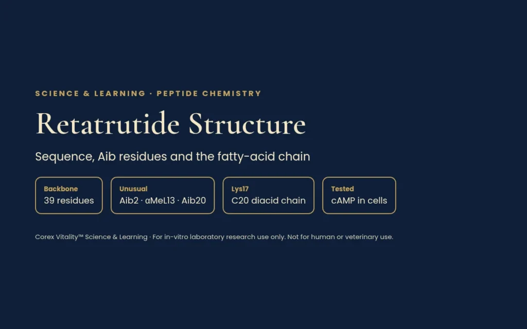
Retatrutide (code name LY3437943) is a synthetic peptide made of 39 building blocks joined in one chain.
Three of those building blocks are unusual: Aib at positions 2 and 20, and α-methyl-leucine at position 13.
A fatty-acid chain (a C20 fatty diacid on a short linker) is fixed to the lysine at position 17.
In cell tests, it switched on three related receptors: the GIP, GLP-1 and glucagon receptors. Cryo-EM images show how it fits into each one.
Our guide to GLP-1, GIP and glucagon receptor peptides explains the receptor family as a whole. This article explains retatrutide structure piece by piece, and how labs measured its activity in cells.
1. First, some basic words
A peptide is a short chain of amino acids, the small building blocks that proteins are made from.
Each amino acid in the chain is called a residue. Residues are numbered from the start of the chain (the N-end) to the finish (the C-end).
The order of residues is the sequence. It is written in one-letter codes, such as Y for tyrosine and K for lysine.
A receptor is a protein on the cell surface. When the right molecule fits it, the cell sends a signal inside. A molecule that switches a receptor on is called an agonist.
2. The 39-residue backbone
Retatrutide is built on a sequence based on GIP (gastric inhibitory polypeptide, a natural gut hormone). The chemical name listed in the PubChem database (CID 171390338) spells out the full chain:
K* is the lysine at position 17 that carries the fatty-acid chain. The NH₂ at the end means the last residue is an amide: the usual acid group at the C-end has been swapped for a slightly different chemical group. PubChem lists the formula as C₂₂₁H₃₄₂N₄₆O₆₈, with a molecular mass of about 4,731.
Schematic of retatrutide: the 39-residue chain, the three unusual residues (gold), the fatty-acid chain on Lys17, and the in-vitro cAMP potency results. Diagram: Corex Vitality™ Science & Learning.
3. The three unusual building blocks
Cells build proteins from 20 standard amino acids. Retatrutide contains three that are not on that list. Chemists call these non-coded amino acids.
Aib (α-aminoisobutyric acid) at positions 2 and 20. It looks like alanine with an extra small carbon group (a methyl group) on its central carbon.
α-methyl-leucine (αMeL) at position 13. This is leucine with the same kind of extra methyl group.
The 2026 review by Mateescu and colleagues notes that Aib at position 2 makes the chain harder for an enzyme called DPP-4 to cut. DPP-4 clips the first two residues off many peptides of this family.
4. The fatty-acid chain on position 17
Adding a fatty chain to a peptide is called lipidation. In retatrutide, the side chain of lysine 17 carries three parts, in this order:
AEEA: one short, flexible spacer (its full name is 8-amino-3,6-dioxaoctanoic acid).
γ-Glu: a gamma-glutamic acid link.
C20 fatty diacid: a straight chain of 20 carbon atoms with an acid group at each end.
The review explains that this chain makes the peptide stick to albumin, a common protein. That matters in the lab, because adding albumin to an assay can change how strong the peptide appears.
5. What the cryo-EM pictures show
Cryo-EM (cryo-electron microscopy) takes thousands of images of frozen molecules and combines them into a 3D model. In 2024, Li and colleagues used it to picture retatrutide bound to each of the three receptors. The resolutions were 2.68, 3.26 and 2.84 ångströms (an ångström is a ten-billionth of a metre).
Residues 1–30 formed one continuous helix, a spiral shape.
Residues 1–13 reached deep into the core of each receptor. Residues 14–30 touched the receptor’s outer parts.
Many contacts were shared by all three receptors. Others were unique to one.
The team then changed single receptor residues and re-tested activity. For example, one change in the GIP receptor (R196Y) cut retatrutide’s potency about 108-fold. The models are public in the Protein Data Bank as 8YW3, 8YW4 and 8YW5.
6. How potency was measured in cells
When these receptors switch on, the cell makes more of a small signal molecule called cAMP (cyclic AMP). Measuring cAMP shows how strongly a peptide activates a receptor.
The original tests used HEK293 cells, a standard human cell line grown in the lab. Each cell line carried just one human receptor, at a low level, so the signal was not over-boosted. Results are given as EC50: the concentration that gives half the maximum response. A lower EC50 means a more potent peptide.
As set out in the 2026 review, retatrutide’s EC50 was 0.0643 nM at the GIP receptor, 0.775 nM at the GLP-1 receptor and 5.79 nM at the glucagon receptor (nM means nanomolar, a very low concentration). Compared with each receptor’s own natural hormone, that is about 8.9 times as potent at GIP, and about 0.4 and 0.3 times as potent at GLP-1 and glucagon.
The review stresses three cautions:
Each ratio compares retatrutide with a different natural hormone, so the three numbers should not be compared directly with each other.
Apparent potency depends on test conditions, such as albumin in the assay, and it differed at mouse receptors.
cAMP is only one readout. At the GLP-1 receptor, retatrutide was a partial agonist for a second signal called β-arrestin recruitment.
7. Making and checking the molecule
Peptides like this are often made by solid-phase peptide synthesis (SPPS), adding one residue at a time to a chain anchored on tiny resin beads. A 2026 paper by Mao and colleagues described another route, where a greasy “tag” helps the growing chain be handled in solution instead.
Labs also need to tell closely related lipidated peptides apart. In 2026, Tong and colleagues validated a liquid chromatography method that separated and identified nine of them, including retatrutide.
The peer-reviewed papers below were used for the science in this article. Each entry was checked against PubMed on 4 November 2026. Summaries describe laboratory, cell-based and structural findings only. Corex Vitality™ is not affiliated with the cited authors, journals or institutions.
Structural insights into the triple agonism at GLP-1R, GIPR and GCGR manifested by retatrutide. Li W, Zhou Q, Cong Z, et al.. Cell Discov. 2024;10(1):77. PMID: 39019866 · DOI: 10.1038/s41421-024-00700-0 Cryo-EM study that solved the structures of retatrutide bound to the GLP-1, GIP and glucagon receptors, with cell-based cAMP tests on mutated receptors to map which contacts matter at each one.
Retatrutide as a Unimolecular GIP/GLP-1/Glucagon Receptor Tri-Agonist: Molecular Design, Pharmaceutical Development and Critical Interpretation of Emerging Phase 3 Evidence. Mateescu DM, Capraru ID, Enache B, et al.. Pharmaceutics. 2026;18(10):1277. PMID: 42942810 · DOI: 10.3390/pharmaceutics18101277 Review whose molecular-design section sets out the 39-residue sequence, the Aib2, α-methyl-leucine13 and Aib20 residues, the Lys17 fatty diacid side chain and the original cell-based cAMP potency data.
A Hydrophobic Tag-Assisted Liquid-Phase Strategy for the Synthesis of Retatrutide. Mao CY, Pang ZJ, Qiao GY, Dong L. Org Lett. 2026;28(23):7486-7490. PMID: 42224238 · DOI: 10.1021/acs.orglett.6c02001 Synthetic chemistry study describing a hydrophobic tag-assisted liquid-phase method for building retatrutide, as an alternative to solid-phase peptide synthesis.
Development and validation of a multiplexed LC-HRMS method for nine GLP-1 receptor agonists and its pharmaceutical application. Tong LH, Leung KK, Hung CT. J Chromatogr A. 2026;1786:467288. PMID: 42603384 · DOI: 10.1016/j.chroma.2026.467288 Analytical chemistry study that developed and validated a liquid chromatography method to separate and identify nine related lipidated peptides, including retatrutide.
All Corex Vitality™ products are supplied strictly for in-vitro laboratory research use only. Not for human or veterinary use. No information here describes use in humans or animals.
Research you can verify.
Corex Vitality™ Research Updates
Get new Learning Hub articles and stock updates. No spam. Unsubscribe any time.
Manage Consent
To provide the best experiences, we use technologies like cookies to store and/or access device information. Consenting to these technologies will allow us to process data such as browsing behaviour or unique IDs on this site. Not consenting or withdrawing consent may adversely affect certain features and functions.
Functional
Always active
The technical storage or access is strictly necessary for the legitimate purpose of enabling the use of a specific service explicitly requested by the subscriber or user, or for the sole purpose of carrying out the transmission of a communication over an electronic communications network.
Preferences
The technical storage or access is necessary for the legitimate purpose of storing preferences that are not requested by the subscriber or user.
Statistics
The technical storage or access that is used exclusively for statistical purposes.The technical storage or access that is used exclusively for anonymous statistical purposes. Without a subpoena, voluntary compliance on the part of your Internet Service Provider, or additional records from a third party, information stored or retrieved for this purpose alone cannot usually be used to identify you.
Marketing
The technical storage or access is required to create user profiles to send advertising, or to track the user on a website or across several websites for similar marketing purposes.
Research-grade peptides supplied exclusively for in-vitro laboratory research, supported by third-party HPLC testing and transparent batch documentation. Not for human or veterinary use.
By entering this website, you confirm that you are 18 years of age or older, are visiting exclusively for in-vitro laboratory research, and agree to our Terms & Conditions and Privacy Policy.