
Cellular senescence explained
Background science. Summarises published literature for educational purposes.
What is cellular senescence?
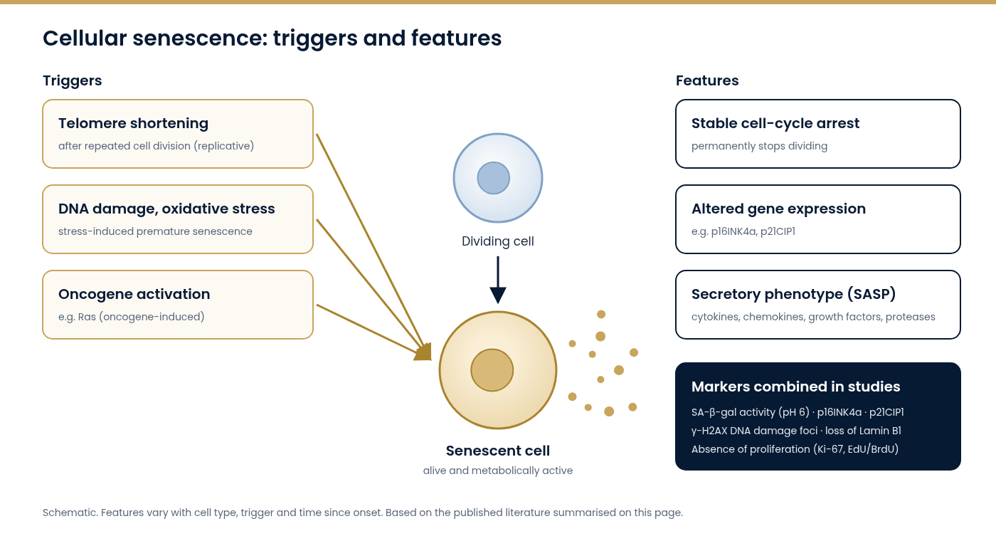
Cellular senescence is a state in which a cell permanently stops dividing but stays alive and metabolically active. Senescent cells differ from quiescent cells, which pause and can divide again, and from cells that die by apoptosis. A consensus paper from researchers in the field describes senescence as a cell state triggered by various stresses and marked by a stable cell-cycle arrest, altered gene expression and a secretory phenotype [1].
A short history
- 1961: the “Hayflick limit”. Leonard Hayflick and Paul Moorhead reported that normal human fibroblasts could divide only a limited number of times in culture before stopping [2].
- 1990: telomeres. Harley and colleagues reported that telomeres (the protective DNA–protein caps at chromosome ends) shortened as human fibroblasts were passaged in culture. This linked replicative limits to telomere length [3].
- 1995: the first widely used marker. Dimri and colleagues described senescence-associated β-galactosidase (SA-β-gal) activity, detectable at pH 6, as a marker of senescent human cells in culture and in skin samples [4].
- 1997: oncogene-induced senescence. Serrano and colleagues showed that an activated oncogene (Ras) could trigger premature senescence, together with accumulation of the proteins p53 and p16INK4a [5].
- 2008: the SASP. Coppé and colleagues characterised the senescence-associated secretory phenotype, the mixture of factors released by senescent cells [6].
Types of senescence studied
| Type | Trigger | Example model |
|---|---|---|
| Replicative senescence | Telomere shortening after repeated cell division | Serially passaged human fibroblasts [2,3] |
| Stress-induced premature senescence | DNA damage (e.g. irradiation, some chemotherapy agents), oxidative stress | Cells treated with genotoxic agents [1] |
| Oncogene-induced senescence | Activation of oncogenes such as Ras | Ras-expressing fibroblasts [5] |
Senescence is heterogeneous: its features depend on the cell type, the trigger and the time since onset [7].
The senescence-associated secretory phenotype (SASP)
Many senescent cells secrete a mixture of cytokines, chemokines, growth factors and proteases, together called the SASP [6]. In experimental systems the SASP has been reported to affect neighbouring cells and tissue environments, and its composition varies between cell types and triggers [7]. Because of this, the SASP is a major subject of research. It is also why some studies investigate senomorphic agents, which change the SASP without removing the cell.
How laboratories detect senescent cells
No single marker is specific to senescence, so the consensus recommendation is to combine several markers [1]. Commonly used markers include:
| Marker | What it indicates | Typical method |
|---|---|---|
| SA-β-gal activity (pH 6) | Increased lysosomal β-galactosidase activity | Histochemical staining [4] |
| p16INK4a | Cyclin-dependent kinase inhibitor; cell-cycle arrest | Immunostaining, qPCR, western blot [1,5] |
| p21CIP1 | Cyclin-dependent kinase inhibitor, downstream of p53 | Immunostaining, western blot [1] |
| Absence of proliferation | Cells not entering S-phase | Ki-67 staining, EdU/BrdU incorporation [1] |
| DNA damage foci | Persistent DNA damage response | γ-H2AX immunofluorescence [1] |
| Loss of Lamin B1 | Altered nuclear envelope | Immunostaining, western blot [1] |
| SASP factors | Secretory phenotype | ELISA, cytokine arrays, RNA-seq [6] |
Senescence and ageing research
The accumulation of senescent cells is listed among the “hallmarks of ageing” in a widely cited framework [8]. In a genetically engineered mouse model, removing p16Ink4a-positive cells delayed several age-associated changes [9]. This finding underpins the senolytics research field (see What are senolytics?). Senescence also has physiological roles reported in development and tissue repair, and it acts as a barrier to the proliferation of damaged cells [1,7]. It is therefore not simply a “harmful” state.
Research use only. This article summarises published scientific literature for educational purposes. It does not describe the effects of any Corex Vitality™ product. Corex Vitality™ products are supplied for laboratory research purposes only and are not intended for human or veterinary use, or for the diagnosis, treatment, prevention or cure of any disease or medical condition.
References
- Gorgoulis V, Adams PD, Alimonti A, et al. Cellular senescence: defining a path forward. Cell. 2019;179(4):813-827. PubMed: https://pubmed.ncbi.nlm.nih.gov/31675495/ · DOI: https://doi.org/10.1016/j.cell.2019.10.005
- Hayflick L, Moorhead PS. The serial cultivation of human diploid cell strains. Exp Cell Res. 1961;25:585-621. PubMed: https://pubmed.ncbi.nlm.nih.gov/13905658/ · DOI: https://doi.org/10.1016/0014-4827(61)90192-6
- Harley CB, Futcher AB, Greider CW. Telomeres shorten during ageing of human fibroblasts. Nature. 1990;345(6274):458-460. PubMed: https://pubmed.ncbi.nlm.nih.gov/2342578/ · DOI: https://doi.org/10.1038/345458a0
- Dimri GP, Lee X, Basile G, et al. A biomarker that identifies senescent human cells in culture and in aging skin in vivo. Proc Natl Acad Sci USA. 1995;92(20):9363-9367. PubMed: https://pubmed.ncbi.nlm.nih.gov/7568133/ · DOI: https://doi.org/10.1073/pnas.92.20.9363
- Serrano M, Lin AW, McCurrach ME, Beach D, Lowe SW. Oncogenic ras provokes premature cell senescence associated with accumulation of p53 and p16INK4a. Cell. 1997;88(5):593-602. PubMed: https://pubmed.ncbi.nlm.nih.gov/9054499/ · DOI: https://doi.org/10.1016/S0092-8674(00)81902-9
- Coppé JP, Patil CK, Rodier F, et al. Senescence-associated secretory phenotypes reveal cell-nonautonomous functions of oncogenic RAS and the p53 tumor suppressor. PLoS Biol. 2008;6(12):2853-2868. PubMed: https://pubmed.ncbi.nlm.nih.gov/19053174/ · DOI: https://doi.org/10.1371/journal.pbio.0060301
- Hernandez-Segura A, Nehme J, Demaria M. Hallmarks of cellular senescence. Trends Cell Biol. 2018;28(6):436-453. PubMed: https://pubmed.ncbi.nlm.nih.gov/29477613/ · DOI: https://doi.org/10.1016/j.tcb.2018.02.001
- López-Otín C, Blasco MA, Partridge L, Serrano M, Kroemer G. Hallmarks of aging: an expanding universe. Cell. 2023;186(2):243-278. PubMed: https://pubmed.ncbi.nlm.nih.gov/36599349/ · DOI: https://doi.org/10.1016/j.cell.2022.11.001
- Baker DJ, Wijshake T, Tchkonia T, et al. Clearance of p16Ink4a-positive senescent cells delays ageing-associated disorders. Nature. 2011;479(7372):232-236. PubMed: https://pubmed.ncbi.nlm.nih.gov/22048312/ · DOI: https://doi.org/10.1038/nature10600