🚚 Free next-day tracked delivery on in-stock orders over £50 🖊️ Peptide Pens → 🖊️ Retatrutide Pens: 20mg £124.99 · 40mg £159.99 → 🎬 View Peptide Videos → 🔬 New: Senolytics & Longevity Research. Explore the science → 👤 Members save 10% on eligible products. Join free → 🚚 Free next-day tracked delivery on in-stock orders over £50 🖊️ Peptide Pens → 🖊️ Retatrutide Pens: 20mg £124.99 · 40mg £159.99 → 🎬 View Peptide Videos → 🔬 New: Senolytics & Longevity Research. Explore the science → 👤 Members save 10% on eligible products. Join free → 🚚 Free next-day tracked delivery on in-stock orders over £50 🖊️ Peptide Pens → 🖊️ Retatrutide Pens: 20mg £124.99 · 40mg £159.99 → 🎬 View Peptide Videos → 🔬 New: Senolytics & Longevity Research. Explore the science → 👤 Members save 10% on eligible products. Join free →
Science & Learning

Peptide Science & Learning

Plain-English guides to peptide chemistry, laboratory testing, storage and published research, all in one place.

Peptide basics

The chemistry, step by step

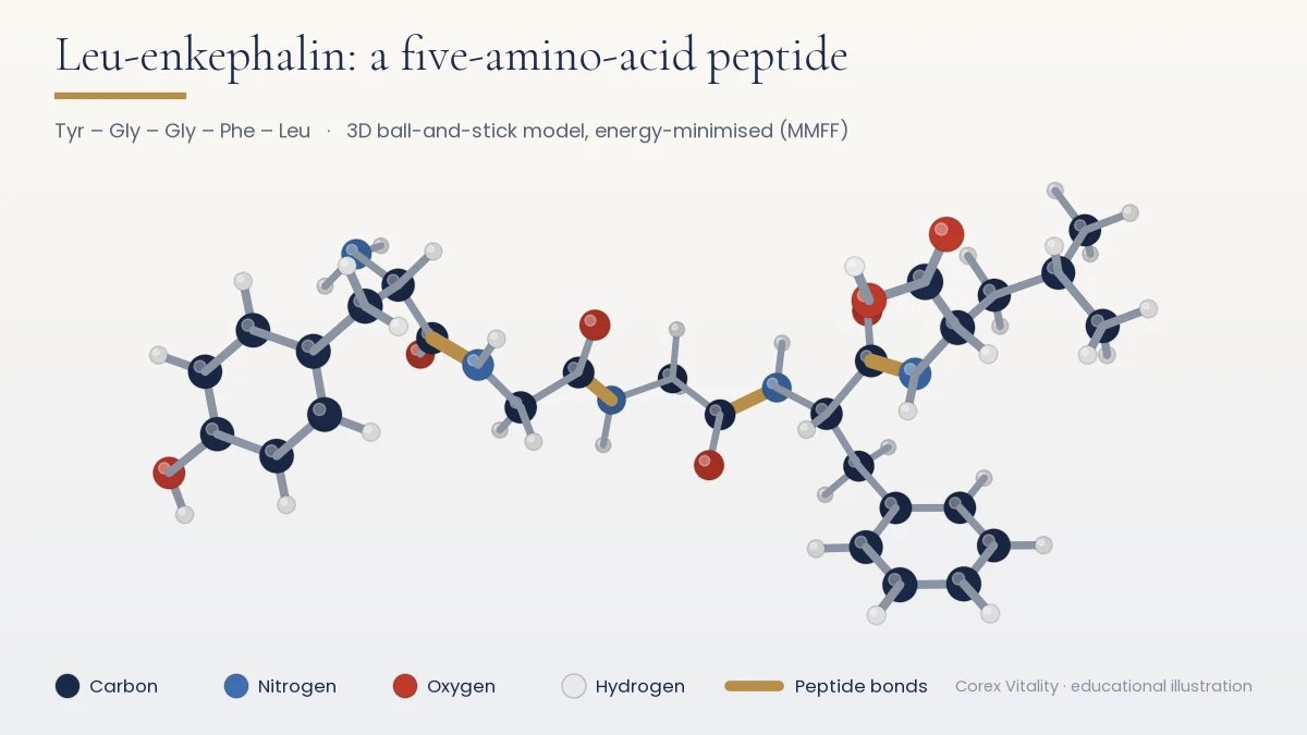
A peptide is a short chain of amino acids, usually 2 to 50, joined by peptide bonds. The order of those amino acids, the sequence, defines exactly which molecule it is. Longer chains are generally called proteins.

Most research peptides are made by solid-phase synthesis, purified by HPLC and freeze-dried into a stable powder.

Storage & handling for research

Keeping research material stable

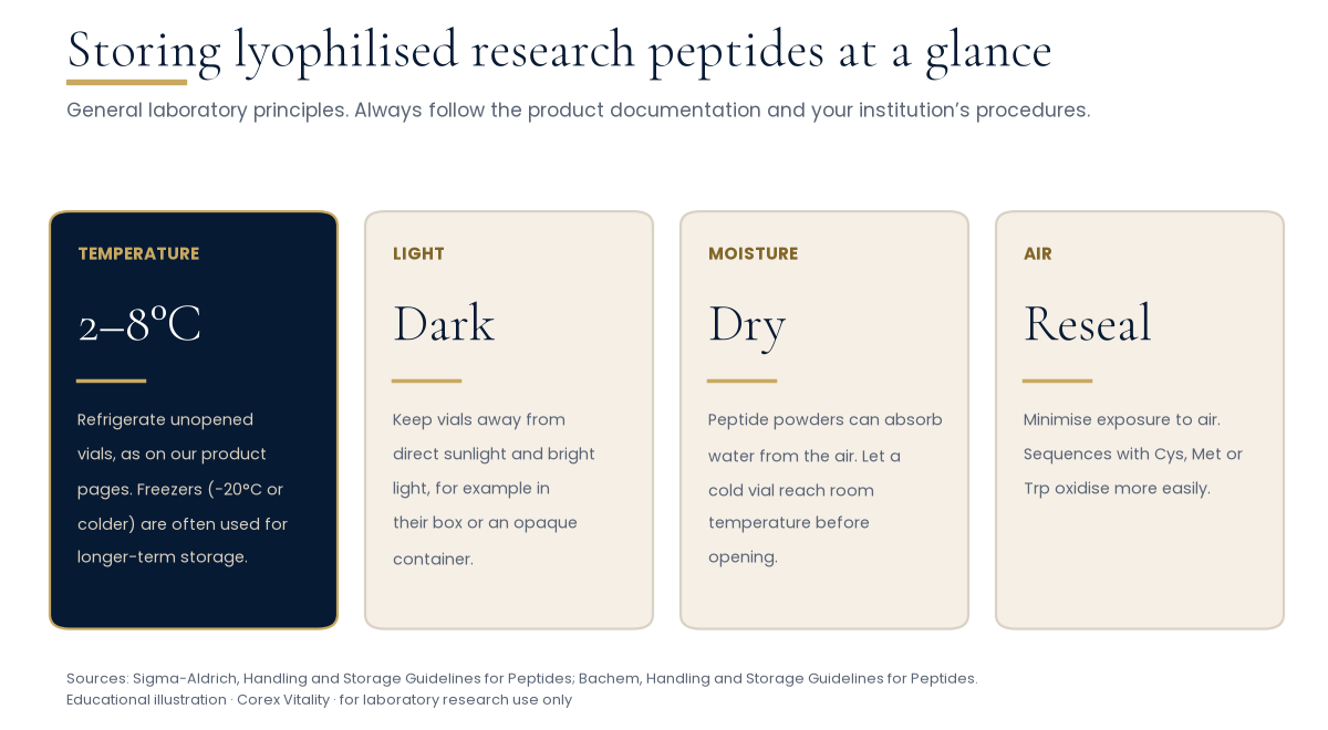
Freeze-dried (lyophilised) peptides are far more stable than solutions, but heat, light, moisture and air can all slowly degrade them.

Four panels on storing lyophilised research peptides: temperature 2 to 8 degrees C for unopened vials with freezers often used long term, keep away from light, keep dry and sealed, and reseal to limit exposure to air
General laboratory principles, summarised from manufacturer handling guides. Current guidance: −20 °C or colder for longer storage of unopened lyophilised vials; 2–8 °C short-term. Product documentation takes priority.

Temperature

For longer storage, keep unopened vials of lyophilised peptide tightly closed at −20 °C or colder. A laboratory fridge (2–8 °C) is fine short-term for vials you plan to open soon, and room-temperature transit is normal for dry peptides. Pre-filled solutions such as pens are the exception: keep them at 2–8 °C and do not freeze.

Once a peptide is in solution, split what you keep into single-use aliquots, freeze them and thaw each only once. Each product page has a Storage & Handling box with compound-specific notes. (Summarised in our own words from Bachem’s handling and storage guidelines.)

Light

Keep vials away from direct sunlight and bright light.

Moisture

Peptide powders are often hygroscopic (they absorb water from the air). Keep vials sealed, and let a cold vial reach room temperature before opening, ideally in a desiccator, so condensation does not form. Work quickly and reseal tightly; absorbed moisture lowers the real peptide content.

Air and records

Reseal promptly; sequences containing cysteine, methionine or tryptophan oxidise more easily. Record each batch number and date received.

Always follow the product documentation and your institution’s own safety and handling procedures. Handle research materials only in a suitable laboratory setting.
Understanding research

How to read a research study

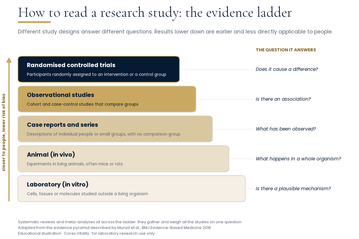
Much published peptide research comes from cell or animal studies. Knowing the type of study tells you what it can, and cannot, show.

Evidence ladder with five rungs from bottom to top: laboratory in vitro studies, animal in vivo studies, case reports and series, observational studies, and randomised controlled trials, each paired with the question it answers
Systematic reviews and meta-analyses sit across the ladder, pooling all the studies on one question. Adapted from Murad et al., BMJ Evidence-Based Medicine, 2016.

Six questions to ask of any study

  1. What was studied?Cells (in vitro), animals (in vivo) or people? Results in cells and mice often do not carry over.
  2. How many?Small samples are more easily swayed by chance. Look for the “n” in each group.
  3. Was there a control group?A fair comparison needs one, ideally assigned at random.
  4. Association or cause?Two things happening together does not mean one caused the other.
  5. Peer-reviewed or preprint?A preprint has not yet been checked by independent experts.
  6. What do the authors say are the limits?The limitations section is often the most useful part of a paper.

Finding the original paper

Summaries, including ours, are a starting point; check the source. PubMed indexes biomedical literature, and a paper’s DOI (digital object identifier) links permanently to the published version.

Read beyond the abstract: the methods show what was actually done, and the results show the size of any difference, not just whether it was statistically significant.

Our research updates set out the model, sample sizes and the limitations the authors noted.

Glossary

Key terms, A to Z by topic

Terms used across our guides. For more ageing-biology terms, see the longevity glossary.

Peptide chemistry

Amino acid
The building block of peptides and proteins: an amino group, a carboxyl group and a distinctive side chain.
Peptide
A short chain of amino acids, typically 2 to 50, linked by peptide bonds.
Peptide bond
The amide link formed when one amino acid’s carboxyl group joins the next one’s amino group, releasing water.
Residue
An amino acid once it is part of a chain.
Sequence
The order of amino acids in a chain, written from the N-terminus to the C-terminus.
N-terminus / C-terminus
The chain ends with a free amino group (N) and a free carboxyl group (C).
Protein
A longer chain, generally above about 50 amino acids, folded into a specific shape.
Dalton (Da)
The unit of molecular mass: one twelfth of the mass of a carbon-12 atom.
SPPS
Solid-phase peptide synthesis: building a chain on resin beads one amino acid at a time (Merrifield, 1963).
Lyophilised
Freeze-dried: frozen, then dried under vacuum, leaving a stable powder.

Testing & certificates

HPLC
High-performance liquid chromatography: separates a sample’s components so each can be measured. Used for purity.
Chromatogram
The HPLC trace: detector signal against retention time, one peak per component.
Retention time
How long a component takes to pass through the column to the detector.
Purity
The main peak’s share of the total HPLC peak area, as a percentage.
Mass spectrometry (MS)
Measures the mass-to-charge ratio (m/z) of charged molecules, to confirm identity.
Batch / lot
Material produced and tested together under one number that links each vial to its certificate.
COA
Certificate of Analysis: the record of tests run on a sample, methods and results.
Endotoxin
Lipopolysaccharide from Gram-negative bacteria, measured with the LAL assay in endotoxin units (EU).

Research methods

In vitro
“In glass”: work on cells, tissues or molecules outside a living organism.
In vivo
Work in a living organism, such as a mouse.
Preclinical
Research before any study in people, mainly in cells and animals.
Randomised controlled trial
Participants are assigned at random to an intervention or a control group.
Peer review
Independent experts assess a paper before a journal publishes it.
Preprint
A paper shared publicly before peer review, for example on bioRxiv.
Systematic review / meta-analysis
A structured search for all studies on one question; a meta-analysis pools their results.

Ageing biology

Cellular senescence
A stable state of cell-cycle arrest in which the cell remains metabolically active.
Senolytic
An agent studied for its ability to selectively induce death of senescent cells.
SASP
Senescence-associated secretory phenotype: signalling molecules released by many senescent cells.
Telomere
The repetitive DNA–protein cap at each chromosome end, which shortens with successive cell divisions in many cell types.
NAD+
Nicotinamide adenine dinucleotide: a coenzyme in redox reactions and a substrate for sirtuins.

Research Use Notice

Corex Vitality™ products are supplied for laboratory research purposes only and are not intended for human or veterinary use, or for the diagnosis, treatment, prevention or cure of any disease or medical condition. Information on this page is general scientific education and does not describe any product’s effects.

Testing & COAs: How to Verify a Peptide COA (UK) · Research range, UK prices & COAs
Compound guides: BPC-157 UK guide: prices & COA · TB-500 UK guide: prices & COA · GHK-Cu UK guide: prices & COA · Retatrutide UK guide: prices & COAs · Semaglutide UK guide: prices & COA · Cagrilintide UK guide: prices & COA · Ipamorelin UK guide: prices & COA · TB-500 + BPC-157 Blend UK guide: prices & COAs · Tirzepatide UK guide: prices & COAs
More guides: Peptide pen storage & COA guide (UK) · Are research peptides legal in the UK? · About Corex Vitality™

Research you can verify.

Corex Vitality™ Research Updates

Get new Learning Hub articles and stock updates. No spam. Unsubscribe any time.

We will send you an email to confirm. Read our privacy policy.

📚 Peer-Reviewed Literature · PubMed Indexed

Published Research — Peer-Reviewed Receptor & Pathway Studies

NCBI PubMed Database ↗ Verify Batch COAs →

Retatrutide (LY3437943)

  1. [1] Li W et al. · Cell Discov (2024)

    Cryo-EM structural determination of retatrutide bound to GLP-1R, GIPR, and GCGR in complex with heterotrimeric Gs proteins, mapping the N-terminal transmembrane recognition contacts underlying triple Class B1 GPCR agonism.

    PMID: 39019866 ↗ DOI: 10.1038/s41421-024-00700-0 ↗
  2. [2] Neumann J et al. · Naunyn Schmiedebergs Arch Pharmacol (2025)

    Ex-vivo pharmacological assay in isolated murine atrial preparations characterizing GCGR-, GIPR-, and GLP-1R-coupled adenylyl cyclase, intracellular cAMP, and protein kinase A (PKA) signalling pathways.

    PMID: 40464942 ↗ DOI: 10.1007/s00210-025-04335-0 ↗
  3. [3] Ma J et al. · Endocrine (2025)

    Pre-clinical murine assay examining renal pro-inflammatory cytokine (TNF-α, caspase-1, NLRP3) and pro-fibrotic marker (fibronectin, α-SMA, collagen I) gene expression alongside serum lipid and butyrate profiles.

    PMID: 39212900 ↗ DOI: 10.1007/s12020-024-03998-8 ↗
  4. [4] Tong LH et al. · J Chromatogr A (2026)

    Analytical validation of a C18 reversed-phase liquid chromatography–high-resolution mass spectrometry (LC-HRMS) method resolving single, dual, and triple receptor agonist peptides with sub-ppm mass accuracy and MS/MS fragment confirmation.

    PMID: 42603384 ↗ DOI: 10.1016/j.chroma.2026.467288 ↗

Tirzepatide (LY3298176)

  1. [1] Willard FS et al. · JCI Insight (2020)

    In-vitro receptor signalling and occupancy study demonstrating full GIPR agonism and biased GLP-1R signalling favouring cyclic AMP (cAMP) generation over β-arrestin recruitment and receptor internalisation.

    PMID: 32730231 ↗ DOI: 10.1172/jci.insight.140532 ↗
  2. [2] Zhao F et al. · Nat Commun (2022)

    Cryo-electron microscopy structure determination of tirzepatide bound to GIPR and GLP-1R, defining the near-atomic extracellular and transmembrane receptor-ligand interactions governing dual incretin agonism.

    PMID: 35217653 ↗ DOI: 10.1038/s41467-022-28683-0 ↗
  3. [3] Sun B et al. · Proc Natl Acad Sci U S A (2022)

    Cryo-EM and molecular dynamics analysis demonstrating how the C20 fatty-diacid acylation moiety and chimeric peptide sequence govern GIPR and GLP-1R receptor activation and reduced GLP-1R desensitisation.

    PMID: 35333651 ↗ DOI: 10.1073/pnas.2116506119 ↗
  4. [4] Regmi A et al. · Cell Metab (2024)

    Cellular and murine receptor-signalling study showing that long-acting GIPR agonism modulates intracellular insulin-receptor cascade cooperativity, glucose transport, glycerol conversion, and lipolytic flux in cultured adipocytes.

    PMID: 38878772 ↗ DOI: 10.1016/j.cmet.2024.05.010 ↗

BPC-157 (Pentadecapeptide)

  1. [1] Chang CH et al. · J Appl Physiol (1985) (2011)

    Ex-vivo rat Achilles tendon explant and cultured tendon fibroblast study demonstrating concentration-dependent acceleration of cellular migration, culture-dish spreading, H2O2 stress survival, and FAK–paxillin phosphorylation.

    PMID: 21030672 ↗ DOI: 10.1152/japplphysiol.00945.2010 ↗
  2. [2] Hsieh MJ et al. · J Mol Med (Berl) (2017)

    In-vitro human vascular endothelial cell and chick chorioallantoic membrane (CAM) study showing upregulated VEGFR2 mRNA/protein expression, receptor internalisation, and VEGFR2–Akt–eNOS phosphorylation.

    PMID: 27847966 ↗ DOI: 10.1007/s00109-016-1488-y ↗
  3. [3] Chang CH et al. · Molecules (2014)

    cDNA microarray, RT-qPCR, and Western blot study in isolated rat Achilles tendon fibroblasts showing concentration- and time-dependent upregulation of growth hormone receptor expression and downstream JAK2 phosphorylation.

    PMID: 25415472 ↗ DOI: 10.3390/molecules191119066 ↗
  4. [4] Huang T et al. · Drug Des Devel Ther (2015)

    In-vitro human umbilical vein endothelial cell (HUVEC) transwell, scratch-migration, and Matrigel tube-formation assays demonstrating concentration-dependent cell-cycle progression and ERK1/2, FAK, and paxillin phosphorylation.

    PMID: 25995620 ↗ DOI: 10.2147/DDDT.S82030 ↗

TB-500

  1. [1] Safer D, Elzinga M, Nachmias VT · J Biol Chem (1991)

    Biochemical study showing that the actin-sequestering peptide Fx and thymosin beta 4 are the same molecule, and describing how it binds actin monomers.

    PMID: 1999398 ↗
  2. [2] Hannappel E · Ann N Y Acad Sci (2007)

    Review of the beta-thymosin peptide family: structure, distribution and known molecular interactions, including actin binding.

    PMID: 17468232 ↗ DOI: 10.1196/annals.1415.018 ↗
  3. [3] Philp D et al. · FASEB J (2003)

    In-vitro endothelial cell adhesion, migration, tube-formation, and chick aortic-ring sprouting study demonstrating that the conserved central 7-amino-acid G-actin-binding motif (LKKTETQ) reproduces the angiogenic signalling of full-length thymosin β4.

    PMID: 14500546 ↗ DOI: 10.1096/fj.03-0121fje ↗
  4. [4] Bock-Marquette I et al. · Nature (2004)

    Embryonic and postnatal cardiomyocyte culture and biochemical study showing that the G-actin-sequestering polypeptide thymosin β4 forms a functional PINCH–integrin-linked kinase (ILK) complex that activates protein kinase B (Akt) signalling.

    PMID: 15565145 ↗ DOI: 10.1038/nature03000 ↗

GHK-Cu (Copper Tripeptide-1)

  1. [1] Pickart L et al. · Nature (1980)

    Foundational biochemical and structure-function study demonstrating that the histidyl-lysyl tripeptide chelates copper(II) in equimolar coordination and facilitates cellular copper transport in cultured hepatoma cells.

    PMID: 7453802 ↗ DOI: 10.1038/288715a0 ↗
  2. [2] Maquart FX et al. · FEBS Lett (1988)

    In-vitro fibroblast culture study showing picomolar-to-nanomolar (10^-12 to 10^-9 M) stimulation of collagen biosynthesis by the GHK–Cu(II) coordination complex independent of cell-number changes.

    PMID: 3169264 ↗ DOI: 10.1016/0014-5793(88)80509-x ↗
  3. [3] Wegrowski Y et al. · Life Sci (1992)

    Radiolabelled [3H]-glucosamine and [35S]-sulfate incorporation assay in cultured human fibroblasts demonstrating biphasic stimulation of extracellular dermatan sulfate and cell-layer heparan sulfate synthesis at 10^-9 to 10^-8 M GHK-Cu.

    PMID: 1522753 ↗ DOI: 10.1016/0024-3205(92)90504-i ↗
  4. [4] Mehr A et al. · Acta Crystallogr D Struct Biol (2020)

    X-ray crystallographic structural study resolving the high-affinity square-pyramidal copper(II) coordination sphere formed by the N-terminal GHK tripeptide motif in recombinant fusion proteins.

    PMID: 33263328 ↗ DOI: 10.1107/S2059798320013741 ↗

NAD+ (Nicotinamide Adenine Dinucleotide)

  1. [1] Ramsey KM et al. · Science (2009)

    Molecular and murine biochemical study demonstrating that nicotinamide phosphoribosyltransferase (NAMPT) and intracellular NAD+ levels oscillate under CLOCK:BMAL1 transcriptional control to regulate SIRT1 deacetylase activity.

    PMID: 19299583 ↗ DOI: 10.1126/science.1171641 ↗
  2. [2] Gomes AP et al. · Cell (2013)

    Cellular and murine bioenergetics study showing that nuclear NAD+ depletion reduces SIRT1 activity and stabilises HIF-1α under normoxic conditions, downregulating mitochondrially encoded oxidative phosphorylation (OXPHOS) subunits.

    PMID: 24360282 ↗ DOI: 10.1016/j.cell.2013.11.037 ↗
  3. [3] Cantó C et al. · Cell Metab (2015)

    Mechanistic review of subcellular NAD+ compartmentalisation, salvage-pathway enzymatic synthesis, sirtuin and PARP cosubstrate consumption, and the mitochondrial unfolded protein response (UPRmt).

    PMID: 26118927 ↗ DOI: 10.1016/j.cmet.2015.05.023 ↗
  4. [4] Camacho-Pereira J et al. · Cell Metab (2016)

    Enzymatic and knockout-mouse study identifying the ectoenzyme NADase CD38 as a primary regulator of cellular NAD+ degradation, mitochondrial oxygen consumption, and SIRT3-dependent protein deacetylation.

    PMID: 27304511 ↗ DOI: 10.1016/j.cmet.2016.05.006 ↗

MOTS-c (Mitochondrial-Derived Peptide)

  1. [1] Lee C et al. · Cell Metab (2015)

    Discovery study identifying a 16-amino-acid peptide encoded within a short open reading frame (sORF) of mitochondrial 12S rRNA that inhibits the folate-methionine cycle, accumulates endogenous AICAR, and activates AMPK in cultured cells.

    PMID: 25738459 ↗ DOI: 10.1016/j.cmet.2015.02.009 ↗
  2. [2] Kim KH et al. · Cell Metab (2018)

    Cell-biology and chromatin-binding study demonstrating AMPK-dependent nuclear translocation of MOTS-c under glucose restriction and direct interaction with ARE-regulated stress transcription factors including NRF2 (NFE2L2).

    PMID: 29983246 ↗ DOI: 10.1016/j.cmet.2018.06.008 ↗
  3. [3] García-Benlloch S et al. · Peptides (2022)

    In-vitro human (LHCN-M2) and murine (C2C12) myoblast culture study showing that wild-type MOTS-c, via its conserved YIFY Src-homology-2 (SH2)-like motif, inhibits IL-6-induced STAT3 transcriptional activity and promotes myotube formation.

    PMID: 35842023 ↗ DOI: 10.1016/j.peptides.2022.170840 ↗
  4. [4] Kumagai H et al. · iScience (2024)

    Cell-free biophysical and murine signalling study identifying casein kinase 2 (CK2) as a direct binding partner of MOTS-c and comparing wild-type vs K14Q variant peptide-protein interaction kinetics.

    PMID: 39559755 ↗ DOI: 10.1016/j.isci.2024.111212 ↗

Semaglutide (Acylated GLP-1 Analogue)

  1. [1] Lau J et al. · J Med Chem (2015)

    Structure-activity relationship study detailing the Aib8 substitution for DPP-4 enzymatic stability and Lys26 C18 fatty-diacid acylation with a γGlu-2xOEG linker for reversible serum albumin binding and high GLP-1R potency.

    PMID: 26308095 ↗ DOI: 10.1021/acs.jmedchem.5b00726 ↗
  2. [2] Knudsen LB et al. · Front Endocrinol (Lausanne) (2019)

    Molecular pharmacology review of acylated GLP-1 analogues examining fatty-acid linker chemistry, non-covalent albumin association, and intracellular GLP-1 receptor cAMP signalling cascades.

    PMID: 31031702 ↗ DOI: 10.3389/fendo.2019.00155 ↗
  3. [3] Malgave A et al. · Eur J Pharm Biopharm (2025)

    Liquid chromatography–high-resolution mass spectrometry (LC-HRMS) preformulation study mapping aqueous peptide bond hydrolysis, deamidation, and buffer-dependent stability profiles of semaglutide across pH and thermal conditions.

    PMID: 40490042 ↗ DOI: 10.1016/j.ejpb.2025.114780 ↗
  4. [4] Liu Y et al. · J Biomol Struct Dyn (2026)

    1.5-microsecond conventional molecular dynamics (cMD) and MM-GBSA free-energy simulation characterising non-covalent binding of the C18 diacid chain of semaglutide across fatty-acid binding pockets on serum albumin.

    PMID: 40720332 ↗ DOI: 10.1080/07391102.2025.2532848 ↗

Cagrilintide (AM833 Lipidated Amylin Analogue)

  1. [1] Kruse T et al. · J Med Chem (2021)

    Medicinal chemistry design and structural optimisation of cagrilintide (AM833), detailing proline substitutions that prevent amyloid fibril formation alongside N-terminal C20 fatty-diacid lipidation for albumin binding.

    PMID: 34288673 ↗ DOI: 10.1021/acs.jmedchem.1c00565 ↗
  2. [2] Fletcher MM et al. · J Pharmacol Exp Ther (2021)

    In-vitro receptor pharmacology study profiling AM833 (cagrilintide) across canonical calcitonin receptor (CTR) and RAMP-complexed amylin receptors (AMY1R, AMY2R, AMY3R), quantifying cAMP, ERK1/2, and β-arrestin signalling.

    PMID: 33727283 ↗ DOI: 10.1124/jpet.121.000567 ↗
  3. [3] Cao J et al. · Science (2022)

    Cryo-electron microscopy determination of calcitonin receptor–RAMP1/2/3 heterodimers bound to peptide agonists, elucidating how RAMP extracellular domains allosterically modulate amylin receptor peptide selectivity.

    PMID: 35324283 ↗ DOI: 10.1126/science.abm9609 ↗
  4. [4] Poulsen C et al. · Mol Pharm (2025)

    Small-angle X-ray scattering (SAXS), circular dichroism, and biophysical characterisation of fatty-acid-acylated amylin analogue self-association, oligomeric equilibrium, and physical stability in aqueous vehicles.

    PMID: 40405715 ↗ DOI: 10.1021/acs.molpharmaceut.5c00161 ↗

CJC-1295 (DAC-Free / Mod GRF 1-29)

  1. [1] Theriault Y et al. · Biopolymers (1988)

    Two-dimensional 1H-NMR spectroscopy and distance-geometry structural determination of the biologically active 29-amino-acid N-terminal domain of growth hormone-releasing factor, identifying two α-helical regions.

    PMID: 3149201 ↗ DOI: 10.1002/bip.360271204 ↗
  2. [2] Stepień T et al. · Neuropeptides (2009)

    In-vitro cell culture study demonstrating specific GHRH(1-29)-NH2 receptor binding, cellular proliferation, and vascular endothelial growth factor (VEGF) secretion in neuroendocrine cell lines.

    PMID: 19747727 ↗ DOI: 10.1016/j.npep.2009.08.005 ↗
  3. [3] Memdouh S et al. · Drug Test Anal (2021)

    Analytical review of synthetic GHRH(1-29) analogues including CJC-1295 and sermorelin, covering solid-phase sequence modifications, proteolytic cleavage pathways, and high-resolution mass spectrometry identification.

    PMID: 34665524 ↗ DOI: 10.1002/dta.3183 ↗
  4. [4] Uçaktürk E et al. · J Pharm Biomed Anal (2026)

    Validation of a nano-LC–quadrupole/Orbitrap high-resolution mass spectrometry workflow for separating and quantifying intact GHRH(1-29) analogues and their N-terminal enzymatic fragments.

    PMID: 41138283 ↗ DOI: 10.1016/j.jpba.2025.117207 ↗

Ipamorelin (Selective GHSR-1a Pentapeptide)

  1. [1] Raun K et al. · Eur J Endocrinol (1998)

    Foundational receptor-pharmacology characterisation of the synthetic pentapeptide ipamorelin (Aib-His-D-2-Nal-D-Phe-Lys-NH2) in primary rat pituitary cells and in-vivo models, demonstrating high GHSR-1a selectivity without ACTH or cortisol elevation.

    PMID: 9849822 ↗ DOI: 10.1530/eje.0.1390552 ↗
  2. [2] Ankersen M et al. · J Med Chem (1998)

    Medicinal chemistry and structure-activity relationship study of ipamorelin pentapeptide analogues evaluating N-methylated backbones and aromatic side-chain conformations in rat pituitary cell assays.

    PMID: 9733495 ↗ DOI: 10.1021/jm9801962 ↗
  3. [3] Johansen PB et al. · Growth Horm IGF Res (1999)

    Pre-clinical rodent study quantifying concentration-dependent longitudinal bone growth rate and somatotroph hormone release following administration of the selective pentapeptide secretagogue ipamorelin.

    PMID: 10373343 ↗ DOI: 10.1054/ghir.1999.9998 ↗
  4. [4] Svensson J et al. · J Endocrinol (2000)

    Dual-energy X-ray absorptiometry (DXA) and peripheral quantitative computed tomography (pQCT) study in rodent models examining cortical and cancellous bone mineral content under GHSR-1a agonist stimulation.

    PMID: 10828840 ↗ DOI: 10.1677/joe.0.1650569 ↗

Epitalon (AEDG Pineal Tetrapeptide)

  1. [1] Khavinson VKh et al. · Bull Exp Biol Med (2003)

    In-vitro telomerase-negative human fetal lung fibroblast (KMST-6) culture study demonstrating induction of hTERT catalytic subunit expression, telomerase enzymatic activity, and telomere length elongation.

    PMID: 12937682 ↗ DOI: 10.1023/a:1025493705728 ↗
  2. [2] Khavinson V et al. · Molecules (2020)

    Immunofluorescence, RT-qPCR, and molecular docking investigation showing that the Ala-Glu-Asp-Gly (AEDG) tetrapeptide binds histone H1/3/6/H4 complexes and upregulates Nestin, GAP43, β-tubulin III, and Doublecortin expression in cultured stem cells.

    PMID: 32019204 ↗ DOI: 10.3390/molecules25030609 ↗
  3. [3] Araj SK et al. · Int J Mol Sci (2025)

    Comprehensive biochemical review of the synthetic tetrapeptide Ala-Glu-Asp-Gly (Epitalon), detailing its telomerase activation pathways, reactive oxygen species scavenging, and epigenetic chromatin interactions in cellular models.

    PMID: 40141333 ↗ DOI: 10.3390/ijms26062691 ↗
  4. [4] Al-Dulaimi S et al. · Biogerontology (2025)

    Comparative in-vitro study across normal epithelial and immortalised cell lines quantifying hTERT mRNA expression, telomerase enzyme activity, and alternative lengthening of telomeres (ALT) promyelocytic leukaemia bodies.

    PMID: 40908429 ↗ DOI: 10.1007/s10522-025-10315-x ↗

SS-31 (Elamipretide / D-Arg-Dmt-Lys-Phe-NH2)

  1. [1] Birk AV et al. · J Am Soc Nephrol (2013)

    Fluorescence spectroscopy and Oxygraph respirometry study demonstrating that the aromatic-cationic tetrapeptide SS-31 binds selectively to inner-mitochondrial-membrane cardiolipin with 1:1 stoichiometry, preventing cytochrome c peroxidase conversion and preserving ATP synthesis.

    PMID: 23813215 ↗ DOI: 10.1681/ASN.2012121216 ↗
  2. [2] Mitchell W et al. · J Biol Chem (2020)

    Biophysical and 31P/2H solid-state NMR study showing that SS-31 partitions into cardiolipin-rich anionic lipid bilayers to down-regulate membrane surface electrostatic potential and prevent divalent calcium-induced lipid packing stress.

    PMID: 32273339 ↗ DOI: 10.1074/jbc.RA119.012094 ↗
  3. [3] Mitchell W et al. · Elife (2022)

    High-resolution structural and biophysical analysis of sixteen SS-31 tetrapeptide variants evaluating aromatic side-chain composition, bilayer insertion depth, membrane surface charge modulation, and cellular ATP production.

    PMID: 35913044 ↗ DOI: 10.7554/eLife.75531 ↗
  4. [4] Campbell MD et al. · Geroscience (2022)

    Quantitative mass-spectrometry phosphoproteomics and mitochondrial respiration study in murine skeletal muscle mapping post-translational phosphorylation changes across electron-transport-chain and metabolic signalling proteins.

    PMID: 36322234 ↗ DOI: 10.1007/s11357-022-00679-0 ↗

FOXO4-DRI (D-Retro-Inverso FOXO4 Peptide)

  1. [1] Baar MP et al. · Cell (2017)

    Foundational study designing the cell-permeable D-retro-inverso peptide FOXO4-DRI, demonstrating competitive disruption of nuclear FOXO4–p53 PML-body complexes, p53 nuclear exclusion, and selective caspase-3/7-mediated apoptosis in senescent cells.

    PMID: 28340339 ↗ DOI: 10.1016/j.cell.2017.02.031 ↗
  2. [2] Huang Y et al. · Front Bioeng Biotechnol (2021)

    In-vitro serial population-doubling culture study demonstrating that FOXO4-DRI selectively clears SA-β-gal-positive senescent chondrocytes and reduces senescence-associated secretory phenotype (SASP) marker expression.

    PMID: 33996787 ↗ DOI: 10.3389/fbioe.2021.677576 ↗
  3. [3] Kim J et al. · FEBS J (2022)

    NMR spectroscopy and isothermal titration calorimetry (ITC) study mapping the molecular binding interface between the FOXO4 forkhead-associated domain and the p53 transactivation domain.

    PMID: 34954873 ↗ DOI: 10.1111/febs.16333 ↗
  4. [4] Bourgeois B et al. · Nat Commun (2025)

    High-resolution NMR, small-angle X-ray scattering (SAXS), and biochemical study demonstrating that FOXO4-DRI directly binds the intrinsically disordered p53 transactivation domain (TAD) with higher affinity than native FOXO4.

    PMID: 40593617 ↗ DOI: 10.1038/s41467-025-60844-9 ↗

KLOW-80 Quad-Blend (GHK-Cu + BPC-157 + TB-500 + KPV)

  1. [1] Chavatte P et al. · J Pharm Pharmacol (2001)

    Two-dimensional 1H-NMR spectroscopy and molecular-mechanics conformational analysis of the α-MSH(11–13) C-terminal tripeptide Ac-Lys-Pro-Val-NH2 (KPV), defining its folded trans-proline secondary structure in solution.

    PMID: 11480545 ↗ DOI: 10.1211/0022357011776360 ↗
  2. [2] Dalmasso G et al. · Gastroenterology (2008)

    In-vitro Caco-2, HT29-Cl.19A, and Jurkat T-cell study demonstrating that the oligopeptide transporter PepT1 mediates intracellular uptake of the KPV tripeptide to inhibit NF-κB and MAPK pro-inflammatory signalling cascades.

    PMID: 18061177 ↗ DOI: 10.1053/j.gastro.2007.10.026 ↗
  3. [3] Land SC · Int J Physiol Pathophysiol Pharmacol (2012)

    In-vitro bronchial epithelial cell study demonstrating that the tripeptide KPV suppresses TNF-α- and respiratory syncytial virus-induced NF-κB nuclear translocation and IL-8/eotaxin chemokine secretion.

    PMID: 22837805 ↗
  4. [4] Songok AC et al. · PLoS One (2018)

    Synthetic peptide chemistry and NMR spectroscopic characterisation of modified Lys-Pro-Val (KPV) tripeptide derivatives engineered to resist proteolytic aminopeptidase degradation in biological matrices.

    PMID: 29953505 ↗ DOI: 10.1371/journal.pone.0199686 ↗

Selank (Thr-Lys-Pro-Arg-Pro-Gly-Pro Tuftsin Analogue)

  1. [1] Kolomin T et al. · Regul Pept (2011)

    PCR-array transcriptional profiling study in murine splenic cells demonstrating that the synthetic heptapeptide Selank (Thr-Lys-Pro-Arg-Pro-Gly-Pro) and its Gly-Pro fragment modulate cytokine, chemokine, and receptor gene expression.

    PMID: 21609736 ↗ DOI: 10.1016/j.regpep.2011.05.001 ↗
  2. [2] Volkova A et al. · Front Pharmacol (2016)

    RT-qPCR gene-expression study in rat frontal cortex examining mRNA levels of 84 neurotransmission-related genes, demonstrating coordinated modulation of GABAA receptor subunits, transporters, and ion-channel transcripts by Selank.

    PMID: 26924987 ↗ DOI: 10.3389/fphar.2016.00031 ↗
  3. [3] Filatova E et al. · Front Pharmacol (2017)

    In-vitro human neuroblastoma IMR-32 cell culture study demonstrating that Selank modulates GABAergic receptor subunit gene expression and acts as an allosteric modulator of GABAA receptor binding.

    PMID: 28293190 ↗ DOI: 10.3389/fphar.2017.00089 ↗
  4. [4] Vyunova TV et al. · Protein Pept Lett (2018)

    Biochemical and receptor-binding study characterising specific high-affinity membrane binding sites for radiolabelled [3H]-Selank and its allosteric modulation of [3H]-GABA binding on rat brain synaptosomal membranes.

    PMID: 30255741 ↗ DOI: 10.2174/0929866525666180925144642 ↗

Semax (Met-Glu-His-Phe-Pro-Gly-Pro ACTH(4-7)PGP)

  1. [1] Dolotov OV et al. · J Neurochem (2006)

    Radioligand binding and immunoassay study using tritium-labelled [3H]-Semax on rat basal forebrain membranes, characterizing specific saturable receptor binding and concentration-dependent elevation of BDNF protein and mRNA in primary glial cultures.

    PMID: 16635254 ↗ DOI: 10.1111/j.1471-4159.2006.03658.x ↗
  2. [2] Zolotarev YA et al. · Amino Acids (2006)

    RP-HPLC and uniformly tritium-labelled peptide degradation study elucidating the sequential aminopeptidase cleavage pathway of Semax (Met-Glu-His-Phe-Pro-Gly-Pro) into stable C-terminal hexapeptide and pentapeptide fragments.

    PMID: 16773243 ↗ DOI: 10.1007/s00726-006-0328-8 ↗
  3. [3] Dolotov OV et al. · Brain Res (2006)

    Quantitative RT-PCR and immunohistochemistry study in rat hippocampal tissue demonstrating rapid upregulation of brain-derived neurotrophic factor (BDNF) and tropomyosin receptor kinase B (TrkB) mRNA and protein levels.

    PMID: 16996037 ↗ DOI: 10.1016/j.brainres.2006.07.108 ↗
  4. [4] Medvedeva EV et al. · BMC Genomics (2014)

    Genome-wide RatRef-12 BeadChip microarray transcriptomic analysis mapping 96 immune-, chemokine-, immunoglobulin-, and vascular-signalling transcripts modulated by the heptapeptide Semax in rat cortical tissue.

    PMID: 24661604 ↗ DOI: 10.1186/1471-2164-15-228 ↗

PT-141 (Bremelanotide / Cyclic Melanocortin Heptapeptide)

  1. [1] Fotsch C et al. · Bioorg Med Chem Lett (2003)

    1H-NMR spectroscopy and distance-restraint conformational study of the cyclic lactam melanocortin agonist scaffold Ac-Nle-cyclo[Asp-Pro-D-Phe-Arg-Trp-Lys] defining the spatial beta-turn orientation of the His/Pro-D-Phe-Arg-Trp pharmacophore.

    PMID: 12824029 ↗ DOI: 10.1016/s0960-894x(03)00412-8 ↗
  2. [2] Sun H et al. · Bioorg Med Chem (2004)

    Nuclear magnetic resonance (NMR) and 3D-QSAR pharmacophore modelling of cyclic lactam heptapeptides derived from the Ac-Nle-cyclo[Asp-His-D-Phe-Arg-Trp-Lys] scaffold, mapping steric and hydrophobic determinants of human MC4R selectivity.

    PMID: 15110848 ↗ DOI: 10.1016/j.bmc.2004.03.017 ↗
  3. [3] Zhang H et al. · Cell Res (2021)

    Cryo-electron microscopy structure determination of the human melanocortin-4 receptor–Gs protein complex bound to cyclic peptide agonists including bremelanotide (PT-141), resolving calcium-cofactor coordination and transmembrane helix 6 activation.

    PMID: 34433901 ↗ DOI: 10.1038/s41422-021-00552-3 ↗
  4. [4] Suzuki S et al. · Anticancer Res (2024)

    In-vitro glioblastoma cell-culture assay investigating melanocortin receptor agonist bremelanotide (PT-141) effects on cellular proliferation, apoptosis markers, and intracellular survivin protein expression.

    PMID: 39197897 ↗ DOI: 10.21873/anticanres.17214 ↗

BAC Water (Sterile Water with 0.9% Benzyl Alcohol Diluent)

  1. [1] Thirumangalathu R et al. · J Pharm Sci (2006)

    Fourier-transform infrared (FTIR), circular dichroism, and size-exclusion chromatography study examining how 0.9% w/v benzyl alcohol preservative interacts with polypeptide tertiary structures across solution pH and storage temperatures.

    PMID: 16729274 ↗ DOI: 10.1002/jps.20619 ↗
  2. [2] Goyal MK et al. · Eur J Pharm Biopharm (2009)

    Size-exclusion chromatography, circular dichroism, differential scanning calorimetry, and dynamic light scattering evaluation of polypeptide stability in aqueous solutions containing bacteriostatic benzyl alcohol preservative.

    PMID: 18929648 ↗ DOI: 10.1016/j.ejpb.2008.09.012 ↗
  3. [3] Bis RL et al. · J Pharm Sci (2015)

    High-sensitivity isothermal titration calorimetry (ITC), intrinsic tryptophan fluorescence, and dynamic light scattering study characterising polypeptide conformational equilibrium in 0.9% benzyl alcohol multi-use aqueous vehicles.

    PMID: 25100180 ↗ DOI: 10.1002/jps.24105 ↗
  4. [4] Zhang J et al. · J Pharm Sci (2015)

    Hydrogen–deuterium exchange mass spectrometry (HDX-MS) analysis mapping local backbone amide exchange kinetics and polypeptide conformational stability in 0.9% benzyl alcohol aqueous buffer systems.

    PMID: 25693946 ↗ DOI: 10.1002/jps.24384 ↗

5-Amino-1MQ (5-Amino-1-Methylquinolinium NNMT Inhibitor)

  1. [1] Babault N et al. · J Med Chem (2018)

    X-ray crystallography and biochemical enzymatic inhibition study characterising nicotinamide N-methyltransferase (NNMT) active-site recognition of S-adenosyl-L-methionine (SAM) and nicotinamide substrate pockets.

    PMID: 29320176 ↗ DOI: 10.1021/acs.jmedchem.7b01422 ↗
  2. [2] Gao Y et al. · J Med Chem (2019)

    Structure-activity relationship, isothermal titration calorimetry, and crystal-structure analysis of small-molecule NNMT inhibitors targeting the nicotinamide and methyl-donor catalytic pocket.

    PMID: 31265285 ↗ DOI: 10.1021/acs.jmedchem.9b00413 ↗
  3. [3] Barrows RD et al. · J Med Chem (2022)

    Enzymatic kinetics, cellular 1-methylnicotinamide (1-MNA) quantification, and structural characterisation of selective small-molecule nicotinamide N-methyltransferase (NNMT) chemical probes.

    PMID: 36288465 ↗ DOI: 10.1021/acs.jmedchem.2c01166 ↗

DSIP (Nonapeptide Research Form: Trp-Ala-Gly-Gly-Asp-Ala-Ser-Gly-Glu)

  1. [1] Monnier M et al. · Experientia (1977)

    Foundational neurochemical and cortical electroencephalography study of the synthetic nonapeptide Trp-Ala-Gly-Gly-Asp-Ala-Ser-Gly-Glu (DSIP) characterizing blood-brain barrier permeability and central delta-wave electrical activity.

    PMID: 590449 ↗ DOI: 10.1007/BF01934026 ↗
  2. [2] Graf M et al. · Peptides (1982)

    Pre-clinical rodent chronobiology study comparing intact synthetic DSIP and its phosphorylated Ser7 analogue (DSIP-P) on circadian locomotor activity rhythms.

    PMID: 6897115 ↗ DOI: 10.1016/0196-9781(82)90161-9 ↗
  3. [3] Nekrasov AN et al. · Biochem Mol Biol Int (1996)

    1H-NMR spectroscopy and molecular-mechanics conformational analysis determining the three-dimensional solution structure and intramolecular hydrogen-bonding network of cyclic DSIP nonapeptide analogues.

    PMID: 8728103 ↗

Kisspeptin-10 (KISS1 C-Terminal Decapeptide YNWNSFGLRF-NH2)

  1. [1] Smith JT et al. · Reproduction (2006)

    Neuroendocrine receptor-signalling study detailing how KISS1-derived C-terminal RF-amide peptides activate the G-protein-coupled receptor GPR54 (KISS1R) on hypothalamic GnRH neurons.

    PMID: 16595713 ↗ DOI: 10.1530/rep.1.00368 ↗
  2. [2] Seminara SB et al. · J Neuroendocrinol (2008)

    Molecular biology and receptor pharmacology review of the KISS1/GPR54 (KISS1R) signalling axis, mapping phospholipase C, intracellular calcium mobilisation, and ERK1/2 phosphorylation downstream of kisspeptin binding.

    PMID: 18601695 ↗ DOI: 10.1111/j.1365-2826.2008.01731.x ↗
  3. [3] Vannecke W et al. · ACS Chem Biol (2017)

    Site-specific furan-oxidation chemical cross-linking and mass-spectrometry study mapping direct ligand–receptor contact residues between the decapeptide Kisspeptin-10 (YNWNSFGLRF-NH2) and living GPR54-expressing cells.

    PMID: 28714670 ↗ DOI: 10.1021/acschembio.7b00396 ↗

AOD-9604 (Tyr-hGH(177–191) Cyclic C-Terminal Peptide Fragment)

  1. [1] Wu Z et al. · Biochem Mol Biol Int (1993)

    Biochemical assay characterising the synthetic disulfide-cyclised C-terminal pentadecapeptide fragment hGH(177–191), demonstrating inhibition of acetyl-CoA carboxylase activity in isolated adipocytes and hepatocytes.

    PMID: 8358331 ↗
  2. [2] Heffernan MA et al. · Am J Physiol Endocrinol Metab (2000)

    Pre-clinical rodent biochemical study examining ex-vivo adipose tissue lipolytic activity, lipogenic enzyme regulation, and absence of IGF-1 or glucose intolerance effects for synthetic C-terminal hGH(177–191) analogues.

    PMID: 10950816 ↗ DOI: 10.1152/ajpendo.2000.279.3.E501 ↗
  3. [3] Ng FM et al. · Horm Res (2000)

    Pre-clinical rodent study of the tyrosine-stabilised hexadecapeptide AOD-9604 (Tyr-hGH(177–191)), characterizing glycerol and free fatty acid release and hepatic pyruvate dehydrogenase activation without hGH receptor competition.

    PMID: 11146367 ↗ DOI: 10.1159/000053183 ↗

SLU-PP-332 (Pan-Estrogen-Related Receptor ERRα/β/γ Agonist)

  1. [1] Billon C et al. · ACS Chem Biol (2023)

    Chemical biology and transcriptomic study characterizing SLU-PP-332 as a selective synthetic pan-ERR (ERRα/β/γ) agonist that upregulates mitochondrial oxidative phosphorylation, FAO, and Ddit4/Slc25a25 transcriptional networks in C2C12 myocytes and murine skeletal muscle.

    PMID: 36988910 ↗ DOI: 10.1021/acschembio.2c00720 ↗
  2. [2] Billon C et al. · J Pharmacol Exp Ther (2024)

    Pre-clinical murine study demonstrating that synthetic ERRα/β/γ activation by SLU-PP-332 increases cellular fatty acid oxidation and mitochondrial respiration rates in skeletal muscle and metabolic tissues.

    PMID: 37739806 ↗ DOI: 10.1124/jpet.123.001733 ↗
  3. [3] Okda HE et al. · Int J Biol Macromol (2026)

    Medicinal chemistry, molecular docking, and structure-activity relationship study of SLU-PP-332 acylhydrazone analogues mapping ligand-binding pocket interactions across estrogen-related receptors ERRα, ERRβ, and ERRγ.

    PMID: 41850449 ↗ DOI: 10.1016/j.ijbiomac.2026.151450 ↗

Tesamorelin (TH9507 Trans-3-Hexenoyl-GHRH(1–44)-NH2)

  1. [1] Tomlinson B · Curr Opin Investig Drugs (2006)

    Pharmacological profile of tesamorelin (TH9507), detailing N-terminal trans-3-hexenoyl conjugation to the 44-amino-acid human GHRH sequence to confer dipeptidyl peptidase-4 (DPP-4) resistance while preserving pituitary GHRH receptor affinity.

    PMID: 17086939 ↗
  2. [2] Wang Y et al. · Expert Opin Investig Drugs (2009)

    Biochemical and endocrine pharmacology review of trans-3-hexenoyl-GHRH(1–44)-NH2 examining pituitary somatotroph receptor activation, pulsatile GH secretion kinetics, and peripheral IGF-1 regulation.

    PMID: 19243281 ↗ DOI: 10.1517/13543780802707658 ↗
  3. [3] Memdouh S et al. · Drug Test Anal (2021)

    Analytical chemistry and liquid chromatography–high-resolution mass spectrometry (LC-HRMS) review covering intact and metabolite detection of tesamorelin, sermorelin, and CJC-1295.

    PMID: 34665524 ↗ DOI: 10.1002/dta.3183 ↗

Sermorelin (GHRH(1–29)-NH2 Synthetic Fragment)

  1. [1] Knoop A et al. · Anal Bioanal Chem (2016)

    Analytical method development combining magnetic-bead immunoaffinity extraction with high-resolution quadrupole-Orbitrap tandem mass spectrometry (LC-HRMS/MS) to identify sermorelin (GHRH 1–29) and related GHRH analogues.

    PMID: 26879649 ↗ DOI: 10.1007/s00216-016-9377-3 ↗
  2. [2] Pont L et al. · J Chromatogr A (2020)

    Comparative analytical chromatography and high-resolution mass spectrometry study optimising antibody-conjugated magnetic bead capture and extraction yield of sermorelin, tesamorelin, and CJC-1295.

    PMID: 32971474 ↗ DOI: 10.1016/j.chroma.2020.461548 ↗
  3. [3] González-López NM et al. · Biomed Chromatogr (2023)

    In-vitro enzymatic and serum stability study mapping proteolytic cleavage kinetics and primary degradation fragments of sermorelin and related somatotropic peptides by LC-MS.

    PMID: 37688464 ↗ DOI: 10.1002/bmc.5741 ↗

Mazdutide (IBI362 / OXM3 Dual GLP-1R/GCGR Agonist)

  1. [1] Gan L et al. · Pharmaceuticals (Basel) (2026)

    Pre-clinical murine and hepatocyte study demonstrating that dual GLP-1R/GCGR agonism by mazdutide downregulates PERK–eIF2α–ATF4 endoplasmic reticulum stress signalling and modulates hepatic lipid-oxidation gene networks.

    PMID: 41901218 ↗ DOI: 10.3390/ph19030371 ↗
  2. [2] Kushner PR et al. · J Am Heart Assoc (2026)

    Translational receptor-signalling review comparing balanced and biased GLP-1R/GCGR dual agonists including mazdutide and survodutide across cardiomyocyte, hepatic, and vascular adenylyl cyclase/cAMP pathways.

    PMID: 42535526 ↗ DOI: 10.1161/JAHA.126.049727 ↗
  3. [3] Qu H et al. · Sci Adv (2026)

    Mechanistic cellular and murine study demonstrating that tubular glucagon receptor (GCGR) activation by dual GLP-1R/GCGR agonists engages vacuolar H+-ATPase (V-ATPase) assembly and autophagic flux.

    PMID: 42555719 ↗ DOI: 10.1126/sciadv.aeg2534 ↗

Survodutide (BI 456906 Acylated Dual GCGR/GLP-1R Agonist)

  1. [1] Thomas L et al. · glycaemic signalling Obes Metab (2024)

    Receptor pharmacology and target-engagement profiling of survodutide (BI 456906), characterizing balanced sub-nanomolar GCGR and GLP-1R potency, C18 diacid albumin binding, and plasma amino-acid/glucagon pharmacodynamic biomarkers.

    PMID: 38560764 ↗ DOI: 10.1111/dom.15551 ↗
  2. [2] Zimmermann T et al. · Mol Metab (2026)

    Fluorescence-labelled peptide biodistribution and whole-brain 3D light-sheet c-Fos imaging study in mice mapping survodutide binding across circumventricular organs (area postrema, median eminence) and parabrachial nuclei.

    PMID: 41638399 ↗ DOI: 10.1016/j.molmet.2026.102326 ↗
  3. [3] Kushner PR et al. · J Am Heart Assoc (2026)

    Comparative mechanistic analysis of dual GCGR/GLP-1R receptor agonists examining receptor-binding stoichiometry, intracellular cAMP generation, and systemic metabolic pathway modulation.

    PMID: 42535526 ↗ DOI: 10.1161/JAHA.126.049727 ↗

Glutathione (GSH / γ-L-Glutamyl-L-Cysteinyl-Glycine Tripeptide)

  1. [1] Zhang H et al. · Semin Cell Dev Biol (2012)

    Biochemical review of intracellular γ-L-glutamyl-L-cysteinyl-glycine (GSH) biosynthesis via glutamate-cysteine ligase (GCL) and glutathione synthetase, detailing protein S-glutathionylation and Nrf2 redox signalling.

    PMID: 22504020 ↗ DOI: 10.1016/j.semcdb.2012.03.017 ↗
  2. [2] Lu SC · Biochim Biophys Acta (2013)

    Molecular enzymology study of the two-step ATP-dependent synthesis of reduced glutathione (GSH), transcriptional regulation of GCLC/GCLM subunits, and thiol-disulfide (GSH/GSSG) redox buffering.

    PMID: 22995213 ↗ DOI: 10.1016/j.bbagen.2012.09.008 ↗
  3. [3] Ferreira MJ et al. · Redox Biol (2023)

    Cellular biochemistry study examining glutathione transport across peroxisomal membranes, glutathione peroxidase/reductase cycles, and hydrogen peroxide detoxification in mammalian organelles.

    PMID: 37804696 ↗ DOI: 10.1016/j.redox.2023.102917 ↗

SNAP-8 (Acetyl Octapeptide-3 / SNAP-25 N-Terminal Mimetic)

  1. [1] Apland JP et al. · J Protein Chem (2003)

    Biochemical and neurochemical assay demonstrating that synthetic oligopeptides mimicking the N-terminal α-helical domain of SNAP-25 competitively inhibit ternary SNARE complex assembly and calcium-dependent vesicular exocytosis.

    PMID: 12760419 ↗ DOI: 10.1023/a:1023423013741 ↗
  2. [2] Li T et al. · Elife (2022)

    Structural and biochemical investigation showing how the N-terminal peptide region of SNAP-25 interacts with vesicular effectors to regulate syntaxin-1A/VAMP2 four-helix bundle SNARE complex formation.

    PMID: 36173100 ↗ DOI: 10.7554/eLife.79926 ↗
  3. [3] Bhasne K et al. · J Biol Chem (2024)

    Biophysical and fluorescence spectroscopy study characterizing the conformational dynamics of unstructured SNAP-25 domains and their assembly into ternary SNARE complexes.

    PMID: 39551143 ↗ DOI: 10.1016/j.jbc.2024.108001 ↗

References are provided for scientific context only. All Corex Vitality™ compounds are supplied strictly for in-vitro laboratory research use (RUO).